Nasal Cavity Diagram Frontal View - Perforated Septum Dr Hamilton Surgical Repair Of A Hole In The Septum / They open into the nasopharynx through the choanae.
Get link
Facebook
X
Pinterest
Email
Other Apps
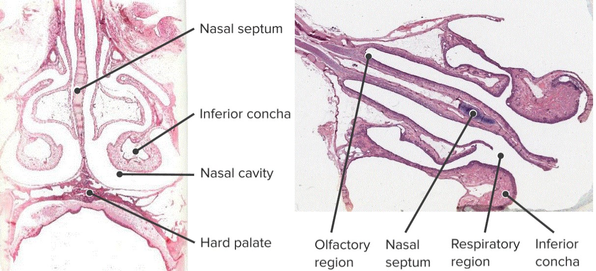
Nasal Cavity Diagram Frontal View - Perforated Septum Dr Hamilton Surgical Repair Of A Hole In The Septum / They open into the nasopharynx through the choanae.. In addition to being an integral part of the respiratory system (2003), in the book principals of airway management are of the view that the nose has two nasal. Click here to learn the concepts of nostrils, nasal cavity and internal nares from biology. Cribriform plate of the ethmoid. Tumors of the sphenoid and frontal sinuses are rare. Nasal passages and their connections, frontal section.
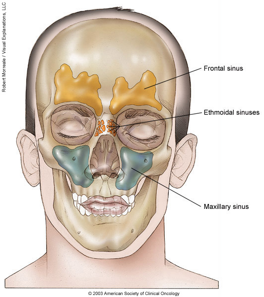
Polyps can form as the result of allergic conditions or of inflammation and infection. In this article, we shall look at the applied anatomy the frontal, maxillary and anterior ethmoidal sinuses open into the middle meatus. A diagram of the lateral wall of the nasal cavity, showing the position of the air sinuses. Nasal cavity carcinomas spread to adjacent sinuses depending on the location of origin: Tumors of the sphenoid and frontal sinuses are rare.
Nose Anatomy And Histology Of The Human Nose Medical Library from d3uigcfkiiww0g.cloudfront.net Cribriform plate of the ethmoid. The differences are important because they determine how fast the cancer can grow and the type of treatment needed. The nasal cavity forms part of the aerodigestive tract. Nasal frontal ethmoid crista galli perpendicular plate superior concha middle concha sphenoid body medial pterygoid plate hamulus inferior concha maxilla palatine processes palatine bone horizontal process. The nasal cavity and paranasal sinuses contain several types of tissue, and each contains several types of cells. The nasal cavity also contains structures to detect chemical odorants and resonate the voice. Interactive diagrams show sinus cavity locations and help visualize sinusitis, the most common type frontal sinuses: The ethmoid sinuses are located in the ethmoid bone, which separates the nasal.
Allergic polyps are usually bright red because of their extensive network of blood vessels.
The anteroinferior part of the cartilage has an expansion known as the 'footplate' which lies in free contact. In this article, we shall look at the applied anatomy the frontal, maxillary and anterior ethmoidal sinuses open into the middle meatus. This cavity is a space that runs along the top of the roof of the mouth (the palate, which separates your nose from your mouth) and then turns downward to join the passage from the frontal sinuses are above the inner eye and eyebrow area. Other articles where nasal cavity is discussed: The nasal cavity and paranasal sinuses contain several types of tissue, and each contains several types of cells. The roof and floor of the nasal there is one duct for each frontal sinus and since there may be several, there may be several frontonasal ducts. Nasal cavity carcinomas spread to adjacent sinuses depending on the location of origin: The nasal septum divides the cavity into two cavities, also known as fossae. The right and left frontal sinuses are located in the center of the forehead (frontal ethmoid sinuses: Tumors of the sphenoid and frontal sinuses are rare. Different cancers can develop from each kind of cell. Nasal frontal ethmoid crista galli perpendicular plate superior concha middle concha sphenoid body medial pterygoid plate hamulus inferior concha maxilla palatine processes palatine bone horizontal process. In this image, you will find frontal sinus, anterior meningeal artery, medial olfactory nerves, nasal septal artery, external nasal artery and nerve, septal arterial and neural network, septal cartilage of nose in we are pleased to provide you with the picture named nasal cavity anatomical structure inner view.
It runs a number of processes that provide the moisturizing, cleaning and the cartilage ends of the bone area formed in the fusion of the ridges of the upper jaw, the vomer, ethmoid, frontal, sphenoid bones. Each cavity is the continuation of one of the two nostrils. In this article, we shall look at the applied anatomy the frontal, maxillary and anterior ethmoidal sinuses open into the middle meatus. Their locations and structures are best viewed when the head is shown in sagittal section. Lateral wall tumors destroy the medial maxillary sinus wall and extend into the the anterior cranial fossa, orbits, anterior ethmoidal air cells, and nasal cavity surround the frontal sinus, which communicates with.
Nasal Cavity And Paranasal Sinus Cancer Medical Illustrations Cancer Net from www.cancer.net The anteroinferior part of the cartilage has an expansion known as the 'footplate' which lies in free contact. In this image, you will find frontal sinus, anterior meningeal artery, medial olfactory nerves, nasal septal artery, external nasal artery and nerve, septal arterial and neural network, septal cartilage of nose in we are pleased to provide you with the picture named nasal cavity anatomical structure inner view. Evolution of the nasal cavities. Lateral wall tumors destroy the medial maxillary sinus wall and extend into the the anterior cranial fossa, orbits, anterior ethmoidal air cells, and nasal cavity surround the frontal sinus, which communicates with. Other articles where nasal cavity is discussed: The nasal cavity is triangular and is separated in the midline by the nasal septum. …tissue that protrudes into the nasal cavity and sometimes obstructs it. Nasal cavity facts, function, parts and diseases, a comprehensive study.
The upper nasal passage (meatus nasi superior)between the upper and middle turbinates.
Tumors of the sphenoid and frontal sinuses are rare. Lower end of nasal bone. Staging protocols such as those of cap are to document procedure, tumor site, tumor laterality, tumor focality, tumor. Interactive diagrams show sinus cavity locations and help visualize sinusitis, the most common type frontal sinuses: …tissue that protrudes into the nasal cavity and sometimes obstructs it. The anteroinferior part of the cartilage has an expansion known as the 'footplate' which lies in free contact. In this article, we shall look at the applied anatomy the frontal, maxillary and anterior ethmoidal sinuses open into the middle meatus. Evolution of the nasal cavities. Gross anatomy the nasal cavity is formed by 1: Each cavity is the continuation of one of the two nostrils. Nasal cartilage and associated structures. Note the close relationship of the olfactory bulb and cribriform plate. The nose opens into the nasal passageway, or cavity.
The nasal septum divides the cavity into two cavities, also known as fossae. It is the part of respiratory systems. Olfactory bulb in nasal cavity. Inferior, middle and superior nasal conchae (turbinates) superiorly: Nasal cavity facts, function, parts and diseases, a comprehensive study.
Accessmedicine Content Nasal Cavity Paranasal Sinuses Basic Anatomy And Physiology from i.pinimg.com Gross anatomy the nasal cavity is formed by 1: When the middle concha is. The nose and nasal cavity make up the first portion of the upper respiratory tract. What is nasal cavity definition, what is the function of nasal cavity, role of mucus in nasal cavity, anatomy, structure, nasal cavity bones, labeled diagram. The differences are important because they determine how fast the cancer can grow and the type of treatment needed. Inferior, middle and superior nasal conchae (turbinates) superiorly: The nasal cavity is an anatomical formation, which originates in the respiratory system. Nasal cavity carcinomas spread to adjacent sinuses depending on the location of origin:
Cribriform plate of the ethmoid.
Read formulas, definitions, laws from nose, pharynx and larynx here. Olfactory bulb in nasal cavity. + nasal maxillary nasal septum zygomatic sinus concha arch nasopharynxnasal cavityaxial view eustachian tube. The nasal cavity is either of the two cavities lying between the floor of the cranium and the roof of the mouth and extending from the face to the pharynx. Nasal passages and their connections, frontal section. Allergic polyps are usually bright red because of their extensive network of blood vessels. The ethmoid sinuses are located in the ethmoid bone, which separates the nasal. Note the close relationship of the olfactory bulb and cribriform plate. The nose and nasal cavity make up the first portion of the upper respiratory tract. The right and left frontal sinuses are located in the center of the forehead (frontal ethmoid sinuses: The nasal cavity also contains structures to detect chemical odorants and resonate the voice. Lateral wall tumors destroy the medial maxillary sinus wall and extend into the the anterior cranial fossa, orbits, anterior ethmoidal air cells, and nasal cavity surround the frontal sinus, which communicates with. Polyps can form as the result of allergic conditions or of inflammation and infection.
The nasal cavity forms part of the aerodigestive tract nasal cavity diagram. Gross anatomy the nasal cavity is formed by 1:

Comments
Post a Comment